


Retina (RTn)

Retina (RTn): Structure, Function, and Clinical Significance
The retina (RTn) is a thin, light-sensitive layer of tissue lining the back of the eye. It serves as the primary sensory interface for vision, converting incoming light into neural signals that are transmitted to the brain via the optic nerve. The retina’s complex structure and microvascular network make it crucial not only for sight but also as a window into systemic and neurological health.
Key Features
Retinal Structure:
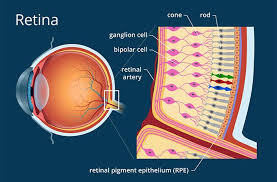
- The retina consists of multiple layers, including photoreceptors (rods and cones), bipolar cells, ganglion cells, and supporting cells.
- The ganglion cell layer (GCL) is particularly important for transmitting visual information to the brain.
Microvascular Network:
- The retina is richly supplied by small blood vessels, including the superficial and deep capillary plexuses.
- These vessels are readily visualized with noninvasive imaging techniques such as optical coherence tomography (OCT) and OCT angiography (OCT-A).
Function:
- Photoreceptors detect light and initiate the visual process.
- Neural circuits within the retina process visual information before it is sent to the brain for interpretation.
Clinical and Research Significance
- Retinal diseases such as diabetic retinopathy, age-related macular degeneration, and retinal vascular occlusions are major causes of vision loss.
Systemic and Neurological Insights:
- Because the retina shares embryological origins and microvascular characteristics with the brain, retinal imaging can reveal early signs of systemic diseases, including hypertension, diabetes, and neurodegenerative disorders.
- Changes in retinal structure or microvasculature are increasingly recognized as biomarkers for cerebral small vessel disease, cognitive impairment, and stroke risk.
Noninvasive Biomarker:
- High-resolution OCT and OCT-A allow for detailed assessment of retinal layers and blood flow, providing a noninvasive method to monitor both ocular and systemic vascular health.
Summary Table
| Aspect | Details |
|---|---|
| Structure | Multi-layered, includes photoreceptors, bipolar cells, ganglion cells, and microvasculature |
| Function | Converts light to neural signals; initiates and processes visual information |
| Clinical Importance | Site of major eye diseases; reflects systemic vascular and neurological health |
| Imaging | OCT and OCT-A enable detailed, noninvasive visualization of retinal layers and vessels |
| Systemic Relevance | Retinal changes serve as biomarkers for diseases such as diabetes, hypertension, and CSVD |
Reference
Kleiner, R.C., et al. (2022). Structural retinal changes in cerebral small vessel disease. Scientific Reports, 12, 9636.
https://doi.org/10.1038/s41598-022-13312-z
The retina’s unique structure and microvascular network not only enable vision but also provide a valuable, noninvasive window into systemic and neurological health, with retinal imaging increasingly used for early detection and monitoring of both ocular and systemic diseases1.