


CT Arthrograms
CT Arthrograms: Definition and Characteristics
What are CT Arthrograms?
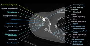
CT arthrograms are advanced imaging procedures that involve the injection of iodinated contrast material directly into a joint, followed by computed tomography (CT) scanning. This method provides high-resolution visualization of intra-articular structures such as the labrum, cartilage, ligaments, and capsule, making it especially useful for detecting joint injuries and subtle abnormalities—most notably in the shoulder and hip.
Characteristics
Mechanism: During a CT arthrogram, contrast is injected into the joint under sterile technique—often using fluoroscopic, ultrasound, or CT guidance. The contrast distends the joint and outlines internal structures, enhancing the visualization of tears, surface irregularities, cartilage defects, and labral pathology. This enables differentiation between normal variants and true injuries, with high diagnostic accuracy for labral tears, articular cartilage loss, and associated soft tissue and bony abnormalities.
Symptoms/Indications: CT arthrograms are typically indicated in cases of persistent joint pain, instability, suspected labral tears, cartilage injury, or following prior surgery when evaluating for complications. They are also especially valuable in patients where MRI is contraindicated or when detailed assessment of bone is required.
Risk Factors: Risks of CT arthrography are minimal but include potential allergic reaction to contrast, infection, temporary post-procedure discomfort, and exposure to ionizing radiation. Patients with iodine allergy or impaired renal function require special consideration.
Clinical Significance
CT arthrograms play a crucial role in diagnosing intra-articular pathology, guiding surgical planning, and assessing unexplained joint pain when standard MRI or CT is inconclusive. Their high spatial resolution and accuracy make them particularly suitable for evaluating labral tears—showing sensitivities up to 97% for hip labrum lesions and excellent correlation with arthroscopic findings. CT arthrography is also valuable in assessing post-surgical joints and in the presence of metallic hardware, where MRI may be limited.
Key Points
- CT arthrograms provide detailed imaging of joint structures after intra-articular injection of contrast.
- They are highly sensitive and specific for detecting labral tears, cartilage injuries, and other intra-articular abnormalities.
- Indicated particularly when MRI is contraindicated or for post-surgical/bony assessment.
- The procedure is generally safe but involves minor risks related to invasive injection and radiation exposure.
Consult with Our Team of Experts Now!
For expert assessment of joint problems and personalized recommendations on imaging—including CT arthrograms—consult with our musculoskeletal radiology team for precise diagnosis and care using Cellular Therapy and Stem Cells.
References:
- Jarraya M, et al. MR-arthrography and CT-arthrography in sports-related shoulder injuries: strengths and limitations. Radiol Med. 2016 Jan;121(1):43-54. doi:10.1007/s11547-015-0581-4. Available at: https://www.ncbi.nlm.nih.gov/pmc/articles/PMC4805613/
- Wyler A, et al. Comparison of MR-arthrography and multidetector-spiral-CT arthrography for the measurement of hip cartilage thickness. Skeletal Radiol. 2009 Jan;38(1):49-56. doi:10.1007/s00256-008-0554-8. Available at: https://pubmed.ncbi.nlm.nih.gov/18614381/
- Magee T. Shoulder MR arthrography: which patient group benefits most? AJR Am J Roentgenol. 2004 Nov;183(4):1077-81. doi:10.2214/ajr.183.4.1830969. Available at: https://ajronline.org/doi/10.2214/ajr.183.4.1830969
- Andreisek G, et al. MR arthrography of the shoulder, hip, and wrist: indications and diagnostic value. Radiographics. 2007;27(6):1619-1654. doi:10.1148/rg.276076066. Available at: https://ajronline.org/doi/10.2214/AJR.06.0719
- Burkhart SS, et al. MR arthrography vs CT arthrography in the evaluation of the glenoid labrum: Diagnostic accuracy and clinical indications. Clin Imaging. 2024 Mar;86:7-14. doi:10.1016/j.clinimag.2024.01.005. Available at: https://doi.org/10.1016/j.clinimag.2024.01.005