


Charcot Joints

Charcot Joints (Neuropathic Arthropathy): Overview, Causes, Pathogenesis, and Management
Definition
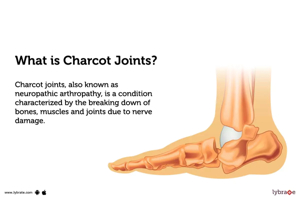
Charcot joint, also known as neuropathic arthropathy or Charcot neuroarthropathy, is a progressive, destructive joint disorder characterized by fragmentation, dislocation, and deformity of bones and joints. It occurs in the setting of peripheral neuropathy, where loss of protective sensation leads to repeated trauma and inflammation unnoticed by the patient.
Etiology
Charcot joints most commonly affect the foot and ankle, especially in patients with diabetic peripheral neuropathy. Other causes include:
- Diabetes mellitus (most common in the U.S.)
- Alcoholic neuropathy
- Spinal cord injury
- Syringomyelia (commonly affects upper extremity joints like the shoulder)
- Neurosyphilis (tabes dorsalis, often affects the knee)
- Leprosy
- Congenital insensitivity to pain
- Other neuropathies (e.g., familial dysautonomia)
Pathogenesis
Two main theories explain Charcot joint development:
- Neurotraumatic Theory:
Loss of sensation and proprioception leads to repetitive microtrauma and joint injury without protective pain feedback. This trauma causes inflammation, bone resorption, and joint destruction. - Neurovascular Theory:
Autonomic neuropathy causes increased blood flow (hyperemia) to the bones, stimulating osteoclastic activity and bone resorption, weakening the bone and predisposing it to fractures and deformity.
Both mechanisms likely contribute to disease progression.
Clinical Presentation
- Early (Acute) Stage:
- Redness, swelling, warmth (3–7°C warmer than contralateral joint)
- Mild to moderate pain (often less than expected)
- Joint instability and deformity begin
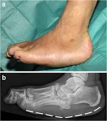
- Radiographs may show bone fragmentation, joint dislocation, and soft tissue swelling.
- Coalescence Stage:
- Inflammation decreases
- Bone healing begins with fusion of fragments
- Swelling and redness subside.
- Reconstruction Stage:
- Bone remodeling and sclerosis occur
- Joint deformity and instability persist
- Foot may develop a rocker-bottom deformity due to collapse of the arch.
Commonly Affected Joints
- Foot and ankle: Most common site, especially tarsometatarsal and metatarsophalangeal joints.
- Knee: Often in neurosyphilis.
- Shoulder: Common in syringomyelia.
- Spine: In spinal cord injury.
- Hip and other joints: Less common.
Diagnosis
- Clinical suspicion in patients with neuropathy presenting with warm, swollen joints.
- Imaging:
- Laboratory tests to exclude infection or inflammatory arthritis.
Management
- Early Recognition: Critical to prevent progression and deformity.
- Offloading and Immobilization: Total contact casting or custom braces to protect the joint and allow healing.
- Patient Education: Avoid weight-bearing on affected limb.
- Surgery: Reserved for severe deformity, instability, or ulceration not responsive to conservative care.
- Monitoring: Regular follow-up to detect complications like ulcers or infection.
Complications
- Joint deformity and instability
- Plantar ulcers due to abnormal pressure points
- Osteomyelitis and infection
- Amputation in severe cases
Summary Table
| Aspect | Details |
|---|---|
| Definition | Progressive joint destruction due to neuropathy-induced loss of protective sensation |
| Common Causes | Diabetes, syringomyelia, neurosyphilis, spinal cord injury, alcoholic neuropathy |
| Pathogenesis | Neurotrauma (repetitive injury) and neurovascular (hyperemia-induced bone resorption) |
| Typical Joints | Foot and ankle (most common), knee, shoulder, spine |
| Clinical Features | Warm, swollen, red joint; mild pain; deformity; rocker-bottom foot in late stages |
| Diagnosis | Clinical exam, X-rays, MRI, exclude infection |
| Treatment | Offloading, immobilization, patient education, surgery if needed |
| Complications | Ulcers, infection, deformity, amputation |
References:
- Petrova NL, Edmonds ME. “Charcot neuro-osteoarthropathy.” Diabetes/Metabolism Research and Reviews. 2016;32 Suppl 1:284-289.
DOI: 10.1002/dmrr.2776
Charcot joint is a serious complication of peripheral neuropathy, especially diabetes, requiring early diagnosis and multidisciplinary management to prevent severe deformity and limb loss.