


Achilles tendon
Achilles Tendon: Overview, Anatomy, and Function
What is the Achilles Tendon?
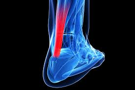
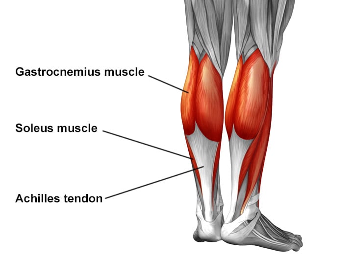
The Achilles tendon, also known as the calcaneal tendon, is the strongest and thickest tendon in the human body. It connects the calf muscles—the gastrocnemius and soleus—to the heel bone (calcaneus). This tendon plays a critical role in enabling movements such as walking, running, and jumping by transmitting the force from the calf muscles to the foot.
Anatomy
Located at the back of the lower leg, the Achilles tendon begins near the middle of the calf and inserts onto the posterior surface of the calcaneus. It receives muscle fibers primarily from the soleus on its inner surface almost to its lower end. The fibers of the tendon spiral approximately 90 degrees on their path to the heel, a design that concentrates stress and enhances force transmission to the foot during movement.
The tendon is surrounded by a paratenon—a connective tissue sheath allowing gliding movement—rather than a true synovial sheath. Its blood supply is relatively poor, primarily from branches of the posterior tibial artery, which contributes to its vulnerability to injury.
Function
The primary function of the Achilles tendon is to facilitate plantar flexion of the foot at the ankle, which means pointing the toes downward. This movement is essential for pushing off during walking, running, and jumping. It also stabilizes the ankle joint during the gait cycle.
During activities such as running, the Achilles tendon can bear loads up to 12 times body weight in young, healthy individuals, making it one of the most heavily stressed tendons in the body. Its elasticity and composition of primarily type I collagen and elastin fibers enable it to absorb shock and efficiently transfer muscular forces.
Clinical Importance
The Achilles tendon is prone to injury, especially tears or ruptures caused by sudden excessive stress or degeneration (tendinopathy). Symptoms of rupture include a sudden audible snap followed by pain and difficulty walking. Treatment depends on the severity, ranging from conservative management with immobilization to surgical repair for complete tears.
Key Points
- The Achilles tendon is the largest and strongest tendon, connecting calf muscles to the heel.
- It enables foot plantar flexion crucial for locomotion and balance.
- Its spiral fiber orientation enhances stress handling and force transmission.
- Poor blood supply makes it susceptible to injury and slow healing.
- Injuries range from tendinopathy to complete rupture, requiring varied treatment approaches.
Consult with Our Team of Experts Now!
For evaluation and management of Achilles tendon health or injuries, consult with our specialist team for comprehensive diagnosis, treatment, and rehabilitation.
References:
Del Buono A, Voight ML, Maffulli N. Achilles tendon: functional anatomy and novel emerging concepts. Muscles Ligaments Tendons J. 2012 Dec 19;2(3):169-177. doi:10.11138/mltj/2012.2.3.169. Available at: https://www.ncbi.nlm.nih.gov/pmc/articles/PMC3609991/