


Pleural Fluid Analysis (PFA)

Pleural Fluid Analysis (PFA):
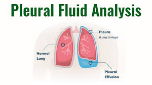
Pleural fluid analysis (PFA) is a key diagnostic tool used to evaluate pleural effusions, especially to distinguish between benign and malignant causes. It involves examining the physical, chemical, cellular, and molecular characteristics of the fluid obtained via thoracentesis.
Key Components of Pleural Fluid Analysis
- Physical Examination:
- Color, clarity, odor, and presence of blood or chyle.
- Biochemical Tests:
- Protein: Differentiates exudate from transudate (Light’s criteria).
- Lactate Dehydrogenase (LDH): Elevated in exudates; used in Light’s criteria.
- pH: Usually low in empyema or malignancy.
- Glucose: Low in infections, rheumatoid pleuritis, or malignancy.
- Amylase: Elevated in esophageal rupture or pancreatic disease.
- Cell Count and Differential:
- Total white blood cell count and differential (neutrophils, lymphocytes, mesothelial cells).
- Malignant effusions often show lymphocytic predominance.
- Cytology:
- Microscopic examination to identify malignant cells.
- Sensitivity around 60%, higher in adenocarcinoma, lower in mesothelioma.
- Repeated sampling increases diagnostic yield (~27% increase with second sample).
- Tumor Markers:
- CEA, CA-125, CA19-9, CYFRA 21-1, and others can support diagnosis, especially when cytology is inconclusive.
- Elevated levels suggest malignancy but are not definitive alone.
- Molecular and Genetic Tests:
- Cell-free DNA methylation analysis and liquid biopsy techniques can improve detection of malignancy, especially when cytology is negative.
- These methods show promise with higher sensitivity and specificity.
- Additional Techniques:
- Cell block preparation: Enhances detection of malignant cells over conventional cytology (positive in up to 96% of cases).
- Immunohistochemistry (IHC): Used on cell blocks for tumor typing (e.g., calretinin, TTF-1).
Diagnostic Approach
- Light’s criteria are used initially to classify effusions as exudates or transudates.
- Cytology remains the mainstay for diagnosing malignant pleural effusions (MPE), with about 60% sensitivity.
- When cytology is negative but suspicion remains high, molecular techniques like liquid biopsy (cell-free DNA methylation) can aid diagnosis.
- Repeat sampling increases diagnostic yield significantly.
Summary of Diagnostic Performance
| Test/Method | Sensitivity | Notes |
|---|---|---|
| Cytology | ~60% | Higher in adenocarcinoma; lower in mesothelioma |
| Cell block analysis | Up to 96% (more sensitive than cytology) | Cost-effective, improves detection |
| Molecular tests (liquid biopsy) | Higher sensitivity (~76%) | Useful when cytology is inconclusive |
| Tumor markers (CEA, CA-125, etc.) | Variable; supportive | Elevated levels suggest malignancy, not definitive |
Conclusion
Pleural fluid analysis is a cornerstone in diagnosing pleural effusions, especially for identifying malignant causes. Combining cytology, tumor markers, cell block techniques, and molecular diagnostics enhances accuracy, enabling early and reliable diagnosis of malignant pleural effusions.
Consult with Our Team of Experts Now!
At DrStemCellsThailand (DRSCT)‘s Anti-Aging and Regenerative Medicine Center of Thailand, we emphasize comprehensive evaluations and personalized treatment plans of Cellular Therapy and Stem Cells for managing various health conditions. If you have questions about Neuromodulation Programs (NMP) or would like more information on our services, consult with our experts today!
Consult with Our Team of Experts Now!
References
- NCBI Bookshelf: Malignant Pleural Effusion
- StatPearls: Malignant Pleural Effusion
- JTS Journal: Diagnosis and Management of Malignant Pleural Effusions
- Nature: Pleural Fluid Analysis in Lung Cancer and Benign Disease
- Nature: Cell-free DNA methylation as a marker of malignancy
- PMC: Diagnostic accuracy of cytology and tumor markers