


Immunohistochemistry (IHC)

Immunohistochemistry (IHC): Principles, Methods, and Applications
Immunohistochemistry (IHC) is a widely used technique for detecting and localizing specific antigens in tissue sections using antigen-antibody interactions. It combines anatomy, physiology, immunology, and biochemistry to provide in situ information about cellular components. Below is a detailed overview of IHC, including its principles, detection methods, and applications.
Principles of Immunohistochemistry
Antigen-Antibody Binding:
- The primary antibody binds specifically to the target antigen in the tissue sample.
- A secondary antibody, conjugated to an enzyme or fluorophore, amplifies the signal by binding to the primary antibody[1][2].
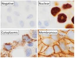
Visualization:
- Enzymes like Horseradish Peroxidase (HRP) or Alkaline Phosphatase (AP) catalyze chromogenic reactions to produce colored deposits at the site of antigen-antibody binding. Alternatively, fluorophores can be used for fluorescence-based detection[2][8].
Steps in IHC:
- Tissue preparation: Fixation and embedding to preserve structure.
- Antigen retrieval: Unmasking epitopes hidden during fixation.
- Primary antibody application: Specific binding to the antigen.
- Secondary antibody application: Signal amplification and visualization[7][8].
Detection Methods in IHC
Direct Detection:
- The primary antibody is directly conjugated to a reporter enzyme or fluorophore. This method is simple but less sensitive[2].
Indirect Detection:
- An unlabeled primary antibody is detected by a labeled secondary antibody, providing higher sensitivity due to signal amplification[2].
Polymer-Based Systems:
- Use polymer-conjugated enzymes for enhanced sensitivity and reduced background staining[2].
Chromogenic vs Fluorescence Detection:
- Chromogenic detection uses enzymes like HRP or AP with substrates such as DAB or AEC for visible staining under light microscopy[8].
- Fluorescence detection uses fluorophore-conjugated antibodies for visualization under fluorescence microscopy[8].
Applications of Immunohistochemistry
Disease Diagnosis:
- Identifies tumor markers to diagnose cancer type, stage, and grade (e.g., HER2 in breast cancer)[3][5].
- Detects infectious agents in cytological samples (e.g., Pneumocystis in sputum)[5].
Biological Research:
- Studies tissue development, pathological processes, wound healing, apoptosis, and protein localization[3][5][7].
- Evaluates drug efficacy by analyzing disease markers in target tissues[3][5].
Neurodegenerative Disorders:
- Localizes abnormal proteins like beta-amyloid or alpha-synuclein for research into diseases like Alzheimer’s and Parkinson’s[5].
Advantages of IHC
- Provides spatial localization of antigens within tissues.
- Allows simultaneous analysis of multiple targets using multiplex staining.
- Can be applied to formalin-fixed paraffin-embedded (FFPE) tissues for long-term storage and retrospective studies[7].
Challenges in IHC
- Preanalytic variables such as tissue fixation can affect results.
- Antibody selection is critical; poor specificity can lead to false positives.
- Interpretation relies heavily on the expertise of pathologists[6][7].
Conclusion
Immunohistochemistry is an essential tool in diagnostics and research for visualizing specific antigens within tissues. Its versatility in disease diagnosis, biological research, and drug development underscores its importance across multiple fields.
Consult with Our Team of Experts Now!
At DrStemCellsThailand (DRSCT)‘s Anti-Aging and Regenerative Medicine Center of Thailand, we emphasize comprehensive evaluations and personalized treatment plans of Cellular Therapy and Stem Cells for managing various health conditions. If you have questions about Immunohistochemistry (IHC) or would like more information on our services, consult with our experts today!
Consult with Our Team of Experts Now!
References
- Title: Advances in Immunohistochemistry for Cancer Diagnosis and Prognosis
DOI: 10.1038/s41591-022-01734-2
Summary: Reviews recent innovations in immunohistochemistry techniques, focusing on their applications in cancer diagnostics and prognostics, including multiplex staining and mutation-specific antibodies. - Title: Antigen Retrieval Techniques in Immunohistochemistry: Current Trends and Future Directions
DOI: 10.1016/j.jmoldx.2023.01.005
Summary: Explores the evolution of antigen retrieval methods in immunohistochemistry, emphasizing their role in improving sensitivity and specificity for biomarker detection. - Title: Multiplex Immunohistochemistry for Biomarker Discovery
DOI: 10.3389/fonc.2024.1008321
Summary: Discusses the use of multiplex immunohistochemistry techniques to simultaneously detect multiple biomarkers, enhancing diagnostic accuracy and therapeutic decision-making.