


![A muscle biopsy is a diagnostic procedure that involves the removal of a small piece of muscle tissue for microscopic examination[3]. It is a valuable tool for diagnosing various neuromuscular disorders, including muscular dystrophy, inflammatory myopathies, metabolic myopathies, and other conditions affecting muscle function[6][4]. The choice of muscle for biopsy depends on the suspected condition and the location of symptoms, but common sites include the biceps (upper arm), deltoid (shoulder), or quadriceps (thigh)[4].](https://cdn.drstemcellsthailand.com/wp-content/uploads/2025/02/image-35.png)
Muscle biopsy (MB)
![A muscle biopsy is a diagnostic procedure that involves the removal of a small piece of muscle tissue for microscopic examination[3]. It is a valuable tool for diagnosing various neuromuscular disorders, including muscular dystrophy, inflammatory myopathies, metabolic myopathies, and other conditions affecting muscle function[6][4]. The choice of muscle for biopsy depends on the suspected condition and the location of symptoms, but common sites include the biceps (upper arm), deltoid (shoulder), or quadriceps (thigh)[4].](https://cdn.drstemcellsthailand.com/wp-content/uploads/2025/02/image-34.png)
Muscle Biopsy: Overview and Diagnostic Significance
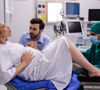
A muscle biopsy (MB) is a diagnostic procedure that involves the removal of a small piece of muscle tissue for microscopic examination[3]. It is a valuable tool for diagnosing various neuromuscular disorders, including muscular dystrophy, inflammatory myopathies, metabolic myopathies, and other conditions affecting muscle function[6][4]. The choice of muscle for biopsy depends on the suspected condition and the location of symptoms, but common sites include the biceps (upper arm), deltoid (shoulder), or quadriceps (thigh)[4].
Purpose of the Muscle Biopsy
The muscle biopsy is performed to:
- Diagnose Neuromuscular Disorders: Identify specific muscle diseases based on structural and biochemical abnormalities[6][4].
- Determine Disease Severity: Assess the extent of muscle damage and inflammation[6].
- Guide Treatment: Provide information to help direct appropriate treatment strategies[6].
Types of Muscle Biopsy Procedures
There are two primary methods for obtaining a muscle biopsy[4][5]:
- Needle Biopsy:
- A specialized needle, approximately 5mm in diameter, is inserted into the muscle to extract a small tissue sample[2][4].
- The procedure is typically brief, lasting less than ten minutes, and is performed under local anesthesia[2][1].
- The small incision is closed with sterile strips and a plaster, requiring no stitches[2][4].
- A potential technique involves using a modified Bergström needle to collect samples from the vastus lateralis muscle[1]. This method involves a small incision, suction, and a cutting blade to extract the tissue[1].
- Open Biopsy:
- A small incision, a few centimeters long (approximately 1.5 inches), is made in the skin to remove a larger section of muscle[2][5].
- The incision is closed with stitches or adhesive strips[2].
- Open biopsies may be performed under local or general anesthesia[4].
- This technique may take about half an hour, but it yields a larger sample, which may be necessary for certain diagnoses[2].
Both types of biopsies have their advantages and disadvantages, and the choice depends on the specific diagnostic needs and the preference of the clinician and patient[2][4].
When to Conduct a Muscle Biopsy
A muscle biopsy may be recommended when patients exhibit symptoms such as[4][5]:
- Muscle weakness
- Muscle pain or cramping
- Muscle stiffness
- Elevated muscle enzymes such as CK/CPK in the blood
Testing Procedure
- Preparation:
- The doctor will explain the procedure, obtain informed consent, and review the patient’s medical history and medications[5].
- Patients should inform their doctor about any medications they are taking, especially blood thinners[5].
- Anesthesia:
- The skin over the biopsy site is cleansed with an antiseptic solution, and a local anesthetic is injected to numb the area[3][1].
- In some cases, an open biopsy may require general anesthesia[4].
- Sample Collection:
- For a needle biopsy, the needle is inserted into the muscle to extract a tissue sample[2][3].
- For an open biopsy, a small incision is made to remove a larger muscle sample[2][3].
- Post-Procedure:
- After the sample is collected, pressure is applied to the site to stop any bleeding[1].
- The incision is closed with sterile strips, stitches, or adhesive strips[2][4].
- The sample is sent to a laboratory for testing and analysis[3].
Interpreting Results
- The muscle tissue is processed and examined under a microscope by a neuropathologist or qualified specialist[6].
- Frozen tissue is often preferred for diagnostic purposes, as it allows for routine histology, enzyme histochemistry, immunostains, and biochemical and molecular studies[6].
- Formalin-fixed tissue is used for routine histological evaluation, special stains, and immunohistochemistry[6].
- Glutaraldehyde-fixed tissue is used for plastic section light microscopy and electron microscopy[6].
- Results are usually available in a few weeks, but more sophisticated analysis may take months[4].
Clinical Implications
- The results of a muscle biopsy can help confirm a diagnosis, determine the severity of the disease, and guide treatment decisions[6][4].
- Muscle biopsies are essential for differentiating between various neuromuscular disorders and identifying specific pathological processes affecting the muscle tissue[6].
Conclusion
A muscle biopsy is a critical diagnostic tool for evaluating neuromuscular disorders. The choice between needle and open biopsy depends on the specific clinical scenario and the need for a larger tissue sample. Proper technique, handling, and analysis of the muscle tissue are essential for accurate diagnosis and appropriate patient management.
Consult with Our Team of Experts Now!
References
- Human Skeletal Muscle Biopsy Procedures Using the Modified. PMC. Link
- Neuromuscular (WERMANS) – muscle biopsy. uhs.nhs.uk. Link
- Muscle biopsy Information | Mount Sinai – New York. mountsinai.org. Link
- Understanding muscle biopsies – Muscular Dystrophy UK. musculardystrophyuk.org. Link
- Muscle Biopsies: What They Are and Why You Might Need One. webmd.com. Link
- What Every Neuropathologist Needs to Know: The Muscle Biopsy. PMC. Link
- Muscle Biopsy: Purpose, Procedure, and Risks – Healthline. healthline.com. Link
- Muscle Biopsy: What It Is, Purpose, Procedure & Recovery. clevelandclinic.org. Link