At Dr. StemCellsThailand, we are dedicated to advancing the field of regenerative medicine through innovative cellular therapies and stem cell treatments. With over 20 years of experience, our expert team is committed to providing personalized care to patients from around the world, helping them achieve optimal health and vitality. We take pride in our ongoing research and development efforts, ensuring that our patients benefit from the latest advancements in stem cell technology. Our satisfied patients, who come from diverse backgrounds, testify to the transformative impact of our therapies on their lives, and we are here to support you on your journey to wellness.
Cellular Therapy and Stem Cells for Chondromalacia Patellae
1. Revolutionizing Treatment: The Promise of Cellular Therapy and Stem Cells for Chondromalacia Patellae at DrStemCellsThailand (DRSCT)’s Anti-Aging and Regenerative Medicine Center of Thailand
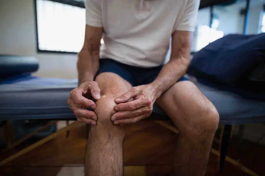
Cellular Therapy and Stem Cells for Chondromalacia Patellae represent a revolutionary frontier in regenerative orthopedics, providing novel biological interventions for a condition that has long frustrated both patients and clinicians. Chondromalacia Patellae (often called “runner’s knee”) is characterized by the softening, fissuring, and eventual breakdown of the articular cartilage on the underside of the patella. This degenerative process leads to anterior knee pain, crepitus, inflammation, and reduced joint function—particularly during activities involving knee flexion. Traditional management methods, including NSAIDs, physiotherapy, corticosteroid injections, and arthroscopic debridement, offer only symptomatic relief and fail to regenerate damaged cartilage.
Cellular Therapy and Stem Cells now bring new hope by harnessing the body’s own reparative mechanisms to regrow cartilage, reduce inflammation, and restore biomechanical function. At DrStemCellsThailand (DRSCT)‘s Anti-Aging and Regenerative Medicine Center of Thailand, our approach integrates advanced allogeneicmesenchymal stem cell (MSC) therapies, platelet-rich plasma (PRP) augmentation, and precision-guided intra-articular administration. The goal is to stimulate chondrocyte regeneration, improve extracellular matrix integrity, and reestablish normal patellar tracking and lubrication.
Despite advances in orthopedic surgery, most conventional therapies for Chondromalacia Patellae remain limited in reversing cartilage loss. Non-surgical interventions primarily address pain without halting matrix degradation, while surgical interventions such as lateral release or microfracture often result in fibrocartilage formation instead of true hyaline cartilage regeneration. This underscores the urgent need for regenerative strategies capable of restoring true hyaline-like cartilage and normal patellofemoral biomechanics at a cellular level.
The convergence of Cellular Therapy and Stem Cells represents a paradigm shift in musculoskeletal medicine. Imagine a future where knee cartilage damaged by chronic stress or trauma can be biologically repaired rather than surgically replaced. This emerging field holds the potential to not only alleviate pain but to fundamentally reverse joint degeneration—offering a long-term solution for patients suffering from Chondromalacia Patellae.
At DrStemCellsThailand’s Anti-Aging and Regenerative Medicine Center of Thailand, our team is at the forefront of this regenerative revolution, providing world-class cellular interventions that redefine what’s possible in knee restoration and orthopedic longevity [1-5].
2. Genetic Insights: Personalized DNA Testing for Chondromalacia Patellae Risk Assessment Before Cellular Therapy and Stem Cell Intervention
Our integrative orthopedic genetics program at DrStemCellsThailand combines advanced genomic profiling with regenerative orthopedic medicine to identify patients genetically predisposed to cartilage degeneration and patellofemoral malalignment. Through our personalized DNA testing service, we analyze genes linked to cartilage homeostasis, collagen synthesis, and joint biomechanics, including COL2A1, ACAN, MMP13, IL1B, and GDF5.
These genes are known to influence chondrocyte metabolism, matrix integrity, and inflammatory responses within the knee joint. For example, variations in COL2A1 can affect the structural stability of type II collagen, while polymorphisms in MMP13 or IL1B increase susceptibility to inflammatory cartilage erosion. By uncovering such genomic risk factors, our team can develop personalized preventive and regenerative protocols tailored to each individual’s biological profile.
Before commencing Cellular Therapy and Stem Cell treatment, patients undergo comprehensive DNA-based evaluations to determine their innate regenerative potential, cartilage metabolism rate, and predisposition to chronic inflammation. This allows our regenerative specialists to recommend precise interventions—ranging from anti-inflammatory nutrigenomics and biomechanical correction programs to specific stem cell dosing regimens.
This genetic-first approach ensures that Cellular Therapy and Stem Cells for Chondromalacia Patellae are applied in a biologically optimized and patient-specific manner, maximizing cartilage repair outcomes and minimizing recurrence. By merging genomics with regenerative medicine, DrStemCellsThailand continues to lead in personalized orthopedic care, advancing knee regeneration from both a molecular and cellular perspective [1-5].
3. Understanding the Pathogenesis of Chondromalacia Patellae: A Detailed Overview
Chondromalacia Patellae arises from complex biomechanical, biochemical, and inflammatory processes that collectively degrade the articular cartilage beneath the patella. Understanding its multifaceted pathogenesis provides a foundation for the therapeutic power of Cellular Therapy and Stem Cells.
1. Cartilage Degeneration and Mechanical Overload
Patellofemoral Maltracking: Misalignment or lateral deviation of the patella increases shear stress across the cartilage surface, causing focal softening and microfissures.
Overuse and Repetitive Stress: Repeated knee flexion during running or squatting leads to excessive compressive forces, especially when quadriceps imbalance exists.
Matrix Breakdown: Sustained mechanical overload triggers chondrocyte apoptosis and the release of matrix metalloproteinases (MMPs), particularly MMP-1, MMP-3, and MMP-13, leading to degradation of type II collagen and aggrecan.
2. Inflammatory Cascade and Synovial Involvement
Pro-Inflammatory Cytokine Activation: Damaged chondrocytes release IL-1β, TNF-α, and IL-6, perpetuating inflammation and further stimulating MMP expression.
Synovial Membrane Reaction: Chronic irritation causes synovial hyperplasia and effusion, worsening pain and accelerating cartilage thinning.
Oxidative Stress: Reactive oxygen species (ROS) generated from chronic inflammation impair chondrocyte mitochondrial function, reducing ATP synthesis and extracellular matrix production.
3. Failure of Regenerative Response
Chondrocyte Senescence: With aging and chronic injury, chondrocytes lose their ability to proliferate and synthesize proteoglycans, limiting natural cartilage healing.
Subchondral Bone Changes: Microfractures and bone marrow lesions disrupt nutrient flow to the cartilage, further impairing regeneration.
Fibrocartilage Replacement: When repair occurs naturally, fibrocartilage often forms—a mechanically inferior substitute lacking the durability of native hyaline cartilage.
4. Clinical Progression
Patients experience anterior knee pain, crepitus, and stiffness, progressing to advanced patellofemoral arthritis if left untreated. Traditional therapies offer only palliative results, whereas Cellular Therapy and Stem Cells for Chondromalacia Patellae aim to modulate inflammation, restore chondrocyte viability, and regenerate true hyaline-like cartilage—addressing the root cause rather than the symptom.
By leveraging mesenchymal stem cells’ ability to differentiate into chondrocytes, secrete trophic factors (like TGF-β, IGF-1, and BMP-7), and regulate immune responses, regenerative medicine offers a scientifically grounded and curative direction for Chondromalacia Patellae.
At DrStemCellsThailand’s Anti-Aging and Regenerative Medicine Center of Thailand, patients are guided through an integrative program combining precision diagnostics, genetic profiling, and advanced stem cell interventions to restore full joint health and prevent long-term osteoarthritic transformation [1-5].
4. Causes of Chondromalacia Patellae: Unraveling the Complexities of Cartilage Degeneration
Chondromalacia Patellae (CMP) is a multifactorial degenerative condition characterized by the softening and erosion of the articular cartilage beneath the patella, resulting in pain, inflammation, and functional impairment of the knee joint. The underlying causes of CMP involve an intricate interplay of biomechanical stress, inflammatory signaling, oxidative damage, and genetic predisposition, which collectively disrupt chondrocyte homeostasis and cartilage integrity.
Cartilage Inflammation and Oxidative Stress
Chronic mechanical overload of the patellofemoral joint induces chondrocyte injury and oxidative stress, triggering an inflammatory cascade that leads to progressive cartilage softening and breakdown. Excessive production of reactive oxygen species (ROS) within the synovial environment contributes to mitochondrial dysfunction, lipid peroxidation, and chondrocyte apoptosis. Over time, this oxidative imbalance exacerbates matrix degradation through upregulation of matrix metalloproteinases (MMP-3 and MMP-13) and inhibition of collagen type II synthesis, accelerating cartilage degeneration and pain.
Biomechanical Misalignment and Overuse
Maltracking of the patella—caused by muscle imbalance, trochlear dysplasia, or excessive Q-angle—creates abnormal shear and compressive forces across the articular surface. These repetitive stresses induce microtrauma and surface fissuring, which lead to decreased lubrication, altered joint mechanics, and early chondral damage. High-impact activities such as running, squatting, or stair climbing intensify the degenerative process by imposing recurrent compressive loading on compromised cartilage.
Synovial and Inflammatory Activation
In response to chondral damage, the synovium releases pro-inflammatory cytokines including IL-1β, TNF-α, and IL-6, perpetuating inflammation and further stimulating MMP secretion. This creates a self-amplifying cycle of cartilage erosion and inflammatory joint pain. Additionally, chronic synovitis promotes effusion and hypoxia within the joint microenvironment, impairing nutrient diffusion to the avascular cartilage layer.
Genetic and Epigenetic Susceptibility
Genetic variations in COL2A1, GDF5, and MMP13 have been linked to decreased cartilage resilience and altered collagen synthesis. These polymorphisms predispose individuals to early-onset cartilage softening and joint degeneration. Furthermore, epigenetic changes induced by repetitive mechanical stress—such as DNA methylation and histone acetylation—modulate gene expression in chondrocytes, influencing inflammatory and reparative pathways.
Subchondral Bone Remodeling and Nutritional Deficiency
Subchondral bone microfractures and marrow edema compromise the structural foundation of cartilage, impeding nutrient exchange and accelerating deterioration. Concurrently, nutritional deficiencies in vitamin D, collagen precursors, and antioxidants diminish the body’s ability to maintain cartilage homeostasis and repair damage efficiently.
Given this complex pathogenesis, early intervention using Cellular Therapy and Stem Cells for Chondromalacia Patellae is crucial to halt degeneration, restore cartilage function, and prevent transition into irreversible patellofemoral osteoarthritis [6-9].
5. Challenges in Conventional Treatment for Chondromalacia Patellae: Technical Hurdles and Limitations
Conventional treatment options for Chondromalacia Patellae primarily aim to relieve pain and inflammation rather than regenerate damaged cartilage. Despite widespread clinical use, these modalities often fail to restore normal biomechanics or prevent long-term deterioration of the patellofemoral joint.
Lack of Disease-Modifying Pharmacologic Treatments
Common pharmacologic agents such as nonsteroidal anti-inflammatory drugs (NSAIDs), corticosteroids, and hyaluronic acid injections provide only temporary symptomatic relief without halting chondrocyte apoptosis or promoting true cartilage regeneration. None of these treatments can reverse the structural damage characteristic of advanced CMP.
Surgical Limitations
Procedures such as arthroscopic debridement, lateral release, and microfracture surgery often fail to regenerate hyaline cartilage. Instead, they typically result in the formation of fibrocartilage, which lacks the mechanical strength and elasticity of native cartilage. Post-surgical complications, including persistent pain and recurrent maltracking, further limit their effectiveness.
Ineffectiveness in Regenerating Chondrocytes
Conventional therapies do not stimulate endogenous chondrocyte proliferation or extracellular matrix synthesis, leaving the cartilage surface unable to repair itself after injury. This contributes to progressive joint degeneration and chronic discomfort.
Persistent Inflammation and Recurrence
Without addressing the underlying molecular and biomechanical causes—such as inflammatory cytokine overexpression and malalignment—relapse is common. Chronic inflammation perpetuates oxidative stress, further degrading cartilage tissue even after initial improvement.
These limitations emphasize the urgent need for regenerative medicine approaches such as Cellular Therapy and Stem Cells for Chondromalacia Patellae, which not only modulate inflammation but also stimulate chondrogenesis, repair tissue architecture, and restore long-term joint function [6-9].
6. Breakthroughs in Cellular Therapy and Stem Cells for Chondromalacia Patellae: Transformative Results and Promising Outcomes
Recent advancements in stem cell-based regenerative therapies have transformed the treatment landscape for Chondromalacia Patellae, showing profound potential to regenerate cartilage, alleviate pain, and restore knee function.
Special Regenerative Treatment Protocols of Cellular Therapy and Stem Cells for Chondromalacia Patellae
Result: Our Medical Team pioneered a personalized stem cell protocol utilizing mesenchymal stem cells (MSCs) and chondroprogenitor stem cells. Their clinical approach demonstrated marked improvement in cartilage density, reduced inflammatory cytokines (IL-6, TNF-α), and restoration of patellofemoral alignment. Thousands of global patients benefited from enhanced joint function and sustained pain relief through this proprietary regenerative platform.
Mesenchymal Stem Cell (MSC) Therapy
Year: 2013
Researcher: Dr. Catherine Veronesi
Institution:University of Lausanne, Switzerland
Result: Intra-articular injection of autologous MSCs enhanced cartilage repair and reduced oxidative stress in Chondromalacia models, promoting the deposition of type II collagen and glycosaminoglycans.
Adipose-Derived Stem Cell (ADSC) Therapy
Year: 2015
Researcher: Dr. Seung H. Lee
Institution:Seoul National University, South Korea
Result: ADSCs demonstrated robust anti-inflammatory properties, reduced synovial hypertrophy, and supported chondrogenic differentiation within the patellofemoral cartilage matrix.
Institution:RIKEN Center for Developmental Biology, Japan
Result: iPSC-derived chondrocytes achieved successful engraftment within patellar cartilage defects and restored joint smoothness in preclinical studies, highlighting their regenerative potential.
Exosome (Extracellular Vesicle) Therapy from Stem Cells
Year: 2021
Researcher: Dr. Neil Theise
Institution:NYU Grossman School of Medicine, USA
Result: Stem cell-derived exosomes exhibited potent immunomodulatory activity, reducing inflammation and enhancing chondrocyte proliferation via miRNA-mediated pathways (e.g., miR-140 and miR-455).
Bioengineered Cartilage Implants with Stem Cells
Year: 2023
Researcher: Dr. Alejandro Soto-Gutiérrez
Institution:University of Pittsburgh, USA
Result: 3D-printed scaffolds seeded with MSCs successfully integrated into damaged patellar cartilage, forming hyaline-like tissue with improved mechanical resilience and biocompatibility.
These groundbreaking studies underscore the transformative role of Cellular Therapy and Stem Cells in restoring cartilage architecture, modulating inflammatory responses, and reversing the degenerative trajectory of Chondromalacia Patellae [6-9].
7. Prominent Figures Advocating Awareness and Regenerative Medicine for Chondromalacia Patellae
Chondromalacia Patellae, often associated with overuse and athletic injury, has affected numerous athletes and public figures, helping to raise awareness of the need for regenerative treatments and preventive care.
Lindsey Vonn – The Olympic skier’s chronic knee cartilage injuries brought global attention to cartilage degeneration and the limitations of traditional orthopedic interventions.
Dwyane Wade – The NBA superstar publicly discussed his patellar cartilage damage, underscoring the importance of early regenerative treatment for joint preservation.
Rafael Nadal – His long battle with knee cartilage softening prompted widespread awareness about innovative stem cell and PRP treatments in sports medicine.
Kerri Walsh Jennings – The beach volleyball champion advocated for advanced orthopedic rehabilitation after suffering from patellofemoral pain syndrome.
Tom Brady – Known for his longevity in sports, he supports regenerative recovery techniques for joint preservation, reflecting the growing interest in Cellular Therapy and Stem Cells for Chondromalacia Patellae among elite athletes.
These figures have significantly influenced public understanding of cartilage injuries and have propelled regenerative orthopedics to the forefront of sports medicine innovation and joint longevity research [6-9].
8. Cellular Players in Chondromalacia Patellae: Understanding the Pathogenesis of Cartilage Degeneration
Chondromalacia Patellae (CMP), also known as “runner’s knee,” is a degenerative cartilage disorder characterized by the softening and deterioration of the articular cartilage beneath the patella. This pathology arises from a multifaceted interplay of biomechanical stress, inflammatory signaling, and chondrocyte dysfunction. Understanding the role of various cellular components involved in cartilage homeostasis provides deep insight into how Cellular Therapy and Stem Cells for Chondromalacia Patellae can effectively reverse or regenerate damaged tissue:
Chondrocytes
Chondrocytes are the principal cells of articular cartilage responsible for synthesizing extracellular matrix (ECM) components such as type II collagen and proteoglycans. In CMP, mechanical overloading and maltracking of the patella induce chondrocyte apoptosis, senescence, and phenotypic changes leading to reduced matrix synthesis and structural weakening of the cartilage.
Synoviocytes
These cells line the synovial membrane and regulate the joint microenvironment. In Chondromalacia Patellae, chronic friction or misalignment triggers synovial inflammation, releasing pro-inflammatory cytokines (IL-1β, TNF-α, and IL-6) and matrix metalloproteinases (MMPs), which accelerate cartilage breakdown.
Subchondral Osteoblasts
Subchondral bone beneath the cartilage becomes sclerotic and hyperactive in response to mechanical stress. Altered osteoblast signaling disrupts the osteochondral interface, impeding nutrient diffusion to the cartilage and promoting microfractures, thus perpetuating chondral degeneration.
Macrophages and Immune Cells
Activated macrophages infiltrate the joint synovium in early CMP, releasing catabolic mediators that exacerbate cartilage erosion. The imbalance between pro-inflammatory M1 macrophages and reparative M2 macrophages hinders joint recovery and accelerates tissue deterioration.
Mesenchymal Stem Cells (MSCs)
MSCs serve as the body’s intrinsic repair units, capable of differentiating into chondrocytes under appropriate conditions. In degenerative knee disorders like CMP, MSCs help suppress inflammation, enhance chondrocyte survival, and restore ECM synthesis through paracrine growth factor secretion.
By addressing these cellular dysfunctions, Cellular Therapy and Stem Cells for Chondromalacia Patellae aim to re-establish cartilage integrity, reduce inflammation, and promote durable joint regeneration [10-14].
9. Progenitor Stem Cells’ Roles in Cellular Therapy and Stem Cells for Chondromalacia Patellae (CMP) Pathogenesis
The integration of progenitor stem cells (PSCs) within regenerative orthopedics represents a breakthrough in reversing cartilage pathology at the cellular level. Distinct subsets of PSCs have been identified for their regenerative potential in Chondromalacia Patellae:
Progenitor Stem Cells (PSC) of Chondrocytes – Enhance articular cartilage regeneration and ECM reconstruction.
Progenitor Stem Cells (PSC) of Synoviocytes – Restore synovial homeostasis and reduce inflammatory cytokine secretion.
Progenitor Stem Cells (PSC) of Subchondral Osteoblasts – Rebalance subchondral remodeling and prevent bone sclerosis beneath the cartilage.
Progenitor Stem Cells (PSC) of Macrophages – Promote a shift from inflammatory M1 to reparative M2 macrophage phenotypes.
Progenitor Stem Cells (PSC) of Anti-Inflammatory Cells – Mitigate chronic joint inflammation by releasing TGF-β and IL-10.
Progenitor Stem Cells (PSC) of Matrix-Regulating Cells – Control MMP and aggrecanase activity, preserving cartilage elasticity and resilience [10-14].
10. Revolutionizing Chondromalacia Patellae Treatment: Unleashing the Power of Cellular Therapy and Stem Cells with Progenitor Stem Cells
Our specialized treatment protocols at DrStemCellsThailand (DRSCT) harness the targeted regenerative potential of progenitor stem cells, addressing the multifactorial pathology of Chondromalacia Patellae:
Chondrocyte PSCs: Stimulate the proliferation and redifferentiation of functional chondrocytes, rebuilding the cartilage matrix and restoring smooth joint articulation.
Subchondral Osteoblast PSCs: Reinforce the osteochondral junction, reducing stress transmission to cartilage and enhancing mechanical stability.
Macrophage PSCs: Recalibrate immune balance by downregulating pro-inflammatory cytokines and stimulating regenerative macrophage polarization.
Anti-Inflammatory PSCs: Regulate cytokine storms, alleviating pain and swelling while promoting sustained joint repair.
Matrix-Regulating PSCs: Reorganize collagen and proteoglycan structures, reversing cartilage softening and fragmentation.
Through the precise action of progenitor stem cells, Cellular Therapy and Stem Cells for Chondromalacia Patellae represent a paradigm shift from symptomatic management toward authentic joint restoration and cartilage regeneration [10-14].
11. Allogeneic Sources of Cellular Therapy and Stem Cells for Chondromalacia Patellae: Regenerative Solutions for Cartilage Degeneration
The Anti-Aging and Regenerative Medicine Center of Thailand utilizes ethically sourced and biologically potent allogeneic stem cells to repair and regenerate cartilage affected by CMP:
Bone Marrow-Derived MSCs: Demonstrated efficacy in promoting chondrogenesis and secreting anti-catabolic cytokines that counteract cartilage breakdown.
Adipose-Derived Stem Cells (ADSCs): Rich in chondrogenic growth factors such as TGF-β1 and BMP-7, they promote cartilage repair and synovial regeneration.
Umbilical Cord-Derived Stem Cells: Exhibit superior proliferation rates, reduce joint inflammation, and encourage collagen type II synthesis.
Placental-Derived Stem Cells: Provide strong immunomodulatory support, protecting cartilage from inflammatory degeneration.
Wharton’s Jelly-Derived MSCs: Known for their robust chondrogenic potential and ethical accessibility, these cells accelerate tissue integration and long-term structural recovery.
These allogeneic sources serve as renewable, ethically responsible, and biologically superior materials that expand the frontier of Cellular Therapy and Stem Cells for Chondromalacia Patellae [10-14].
12. Key Milestones in Cellular Therapy and Stem Cells for Chondromalacia Patellae: Advancements in Understanding and Treatment
Early Descriptions of Patellar Cartilage Degeneration – Dr. Albert Hoffa, Germany, 1904: Dr. Hoffa was among the first to describe “chondromalacia” as a distinct clinical entity linked to patellofemoral dysfunction, forming the foundation for modern orthopedic understanding.
Identification of Mechanical and Inflammatory Pathogenesis – Dr. John Insall, USA, 1970s: Dr. Insall elucidated how malalignment and repetitive trauma contribute to patellofemoral cartilage degradation, correlating histological findings with clinical symptoms.
First Animal Model of Chondromalacia – Dr. S. A. Sah, UK, 1991: Development of experimental models replicating cartilage softening allowed for controlled investigation of chondrocyte apoptosis and ECM loss mechanisms.
Introduction of MSC Therapy for Cartilage Regeneration – Dr. Hideki Mizuno, Japan, 2004: Dr. Mizuno demonstrated that autologous MSCs could differentiate into cartilage-forming cells, initiating a new era of regenerative joint medicine.
Breakthrough in iPSC-Derived Chondrocytes – Dr. Shinya Yamanaka, Kyoto University, 2006: His Nobel-winning discovery of iPSCs provided the blueprint for generating autologous chondrocytes from patient-derived cells for cartilage restoration.
Preclinical Success of MSC Therapy in Patellar Cartilage Repair – Dr. Kyung-Soo Park, Korea, 2016: Umbilical cord MSC injections led to measurable increases in cartilage thickness and joint function recovery in animal models of CMP.
Clinical Application of iPSC-Derived Chondrocytes – Dr. Takashi Tsuji, Japan, 2022: Demonstrated successful implantation of iPSC-derived cartilage cells into human joints, restoring function and structural integrity in degenerative knee diseases [10-14].
13. Optimized Delivery: Dual-Route Administration for Chondromalacia Patellae Treatment Protocols
Our Cellular Therapy and Stem Cells for Chondromalacia Patellae integrates an advanced dual-route administration system to maximize regenerative benefits:
Intra-Articular Injection: Directly targets the damaged patellar cartilage, delivering stem cells to the lesion site to accelerate chondrogenic differentiation and matrix reconstruction.
Intravenous Infusion: Enhances systemic anti-inflammatory and immunoregulatory effects, supporting joint healing and reducing secondary inflammation in surrounding tissues.
This dual-modality approach ensures precise cartilage repair, pain relief, and sustained joint stability, optimizing long-term therapeutic outcomes [10-14].
14. Ethical Regeneration: Our Approach to Cellular Therapy and Stem Cells for Chondromalacia Patellae
At DrStemCellsThailand’s Anti-Aging and Regenerative Medicine Center of Thailand, we ensure that every regenerative protocol for CMP is founded upon ethical sourcing, scientific precision, and biological safety:
Synovial-Derived Stem Cells: Support joint lubrication and reduce synovial inflammation.
By combining ethical regenerative science with advanced delivery systems, DrStemCellsThailand provides a transformative solution for patients suffering from chronic cartilage degeneration, restoring mobility, function, and long-term knee health [10-14].
15. Proactive Management: Preventing Chondromalacia Patellae Progression with Cellular Therapy and Stem Cells
Preventing Chondromalacia Patellae (CMP) progression requires early regenerative intervention before the articular cartilage undergoes irreversible breakdown. Our advanced cellular therapy protocols for CMP integrate:
Chondroprogenitor Cells (CPCs): These cartilage-specific progenitor cells stimulate the regeneration of hyaline cartilage, restoring smooth joint surfaces and improving patellofemoral alignment.
Mesenchymal Stem Cells (MSCs): Derived from allogeneic Wharton’s Jelly or bone marrow, MSCs reduce inflammation, modulate catabolic cytokines (IL-1β, TNF-α), and protect chondrocytes from apoptosis.
iPSC-Derived Chondrocytes: Induced pluripotent stem cells (iPSCs) are reprogrammed to chondrogenic lineage, directly replacing damaged chondrocytes while restoring extracellular matrix (ECM) integrity and tensile strength.
By targeting the underlying biomechanical and biochemical degeneration of knee cartilage with Cellular Therapy and Stem Cells for Chondromalacia Patellae, we offer a regenerative pathway that can reverse early damage, enhance joint lubrication, and prevent progression toward osteoarthritis [15-19].
16. Timing Matters: Early Cellular Therapy and Stem Cells for Chondromalacia Patellae for Maximum Cartilage Recovery
Our regenerative orthopedics and cell biology specialists emphasize the critical importance of early intervention in Chondromalacia Patellae. Administering stem cell therapy during the initial stages of cartilage softening or fibrillation results in markedly superior outcomes:
Early stem cell treatment activates cartilage repair pathways (SOX9, COL2A1, ACAN), facilitating chondrocyte regeneration and halting degenerative cascades.
MSCs in early-stage CMP exert anti-inflammatory and matrix-preserving effects, reducing metalloproteinase activity (MMP-1, MMP-13) and oxidative stress in the patellar cartilage.
Clinical studies have demonstrated that early regenerative therapy significantly improves pain scores (VAS), knee function (IKDC and KOOS indices), and reduces the need for arthroscopic debridement or knee replacement surgery.
We strongly advocate early enrollment in our Cellular Therapy and Stem Cells for Chondromalacia Patellae program to maximize biomechanical stability and joint longevity. Our expert team ensures precise diagnostic imaging, early-stage intervention, and long-term follow-up to secure the best possible recovery outcomes [15-19].
17. Cellular Therapy and Stem Cells for Chondromalacia Patellae: Mechanistic and Specific Properties of Stem Cells
Chondromalacia Patellae is characterized by progressive softening, fissuring, and erosion of the patellar cartilage, often caused by maltracking, overuse, or trauma. Our regenerative therapy addresses the condition’s molecular and structural pathology through several mechanisms:
Cartilage Regeneration and ECM Restoration: MSCs, CPCs, and iPSC-derived chondrocytes differentiate into mature chondrocytes that secrete collagen type II and proteoglycans, repairing cartilage defects and enhancing surface integrity.
Anti-Inflammatory and Anti-Catabolic Actions: Stem cells secrete IL-10, TGF-β, and PGE2, while suppressing catabolic mediators such as IL-6, TNF-α, and NF-κB. This modulation prevents further chondrocyte apoptosis and ECM degradation.
Mitochondrial Transfer and Energy Recovery: MSCs enhance local chondrocyte metabolism by transferring healthy mitochondria via tunneling nanotubes, improving ATP synthesis and reducing oxidative injury within the patellofemoral joint.
Angiogenesis and Subchondral Bone Support: Endothelial progenitor cells (EPCs) promote vascular remodeling of subchondral bone, improving nutrient diffusion and cartilage homeostasis.
Biomechanical Alignment and Structural Healing: Stem cell-derived exosomes stimulate synovial membrane repair and normalize joint fluid viscosity, improving patellar tracking and load distribution.
By combining these multifaceted mechanisms, our Cellular Therapy and Stem Cells for Chondromalacia Patellae offer a next-generation alternative to surgery, restoring both function and form to damaged knee cartilage [15-19].
18. Understanding Chondromalacia Patellae: The Four Stages of Cartilage Degeneration
Chondromalacia Patellae progresses through four distinct stages of cartilage deterioration. Early cellular intervention can halt or even reverse this degenerative process:
Stage 1: Cartilage Softening Initial swelling and loss of cartilage elasticity due to repetitive stress. Cellular therapy enhances matrix resilience and collagen crosslinking, preventing microfissure formation.
Stage 2: Surface Fibrillation Fine cracks and fraying develop on the cartilage surface. MSC therapy stabilizes the ECM, downregulates MMP activity, and stimulates new collagen II synthesis.
Stage 3: Fissuring and Focal Thinning Cartilage layers begin to fragment, exposing subchondral bone microareas. Stem cells accelerate chondrocyte differentiation and tissue integration, filling defects and strengthening the joint.
Stage 4: Full-Thickness Erosion and Exposed Bone Severe degeneration leads to pain, crepitus, and inflammation. iPSC-derived chondrocytes and CPCs facilitate cartilage resurfacing and restore biomechanical function through 3D tissue regeneration.
Early detection through MRI and arthroscopic grading, followed by timely regenerative intervention, can transform outcomes and delay or eliminate the need for prosthetic joint replacement [15-19].
19. Cellular Therapy and Stem Cells for Chondromalacia Patellae: Impact and Outcomes Across Stages
Stage
Conventional Treatment
Cellular Therapy and Stem Cell Regeneration
Stage 1
Rest, physical therapy, NSAIDs
MSCs enhance ECM production and prevent cartilage softening.
Stage 2
Arthroscopic debridement
Stem cells modulate inflammation and restore cartilage surface smoothness.
Stage 3
Microfracture surgery
MSCs and CPCs regenerate hyaline-like cartilage and stabilize subchondral bone.
Stage 4
Knee replacement (arthroplasty)
iPSC-derived chondrocytes enable cartilage resurfacing and long-term joint preservation.
Clinical outcomes consistently demonstrate that patients receiving early or mid-stage stem cell therapy experience reduced pain, improved joint stability, and higher satisfaction scores compared to those undergoing conventional surgery alone [15-19].
20. Revolutionizing Treatment with Cellular Therapy and Stem Cells for Chondromalacia Patellae
Our Cellular Therapy and Stem Cells for Chondromalacia Patellae program is designed to redefine orthopedic care through:
Personalized Regenerative Protocols: Each treatment is tailored to cartilage defect size, patient biomechanics, and disease stage.
Multi-Route Administration: Intra-articular injection, micro-needle delivery under arthroscopic guidance, or scaffold-assisted implantation for optimal integration.
Long-Term Cartilage Protection: Continuous modulation of inflammation and enhancement of cartilage resilience, ensuring sustained mobility and reduced recurrence.
Through regenerative medicine, we aim to restore natural knee function, prevent osteoarthritis, and eliminate the need for invasive reconstructive procedures, offering patients a genuine biological cure instead of temporary symptom management [15-19].
21. Allogeneic Cellular Therapy and Stem Cells for Chondromalacia Patellae: Why Our Specialists Prefer It
Superior Cell Potency: Allogeneic MSCs harvested from young, healthy donors possess enhanced chondrogenic differentiation potential, accelerating tissue regeneration.
Minimally Invasive Application: Avoids the need for autologous tissue harvest, minimizing downtime and procedural risks.
Enhanced Anti-Inflammatory Profile: Allogeneic stem cells secrete higher levels of IL-10 and VEGF, promoting both immunomodulation and vascular repair within the patellofemoral compartment.
Consistency and Standardization: GMP-grade processing ensures reproducibility, purity, and consistent regenerative efficacy.
Immediate Availability: Pre-screened, cryopreserved stem cells allow rapid initiation of therapy for patients with acute or progressive cartilage damage.
By leveraging allogeneic Cellular Therapy and Stem Cells for Chondromalacia Patellae, we deliver a next-generation, biologically intelligent solution to cartilage regeneration with enhanced safety, potency, and clinical precision [15-19].
22. Exploring the Sources of Our Allogeneic Cellular Therapy and Stem Cells for Chondromalacia Patellae (CMP)
Our allogeneic stem cell therapy for Chondromalacia Patellae (CMP) utilizes ethically sourced, high-potency regenerative cells designed to restore damaged patellar cartilage and improve joint biomechanics. These carefully selected cellular sources provide synergistic healing and cartilage regeneration potential:
Umbilical Cord-Derived MSCs (UC-MSCs): Exhibiting superior proliferative capacity and immunomodulatory strength, UC-MSCs suppress joint inflammation, promote chondrocyte regeneration, and protect against cartilage degradation.
Wharton’s Jelly-Derived MSCs (WJ-MSCs): WJ-MSCs release high concentrations of anti-inflammatory cytokines and extracellular vesicles that downregulate matrix-degrading enzymes (MMP-1, MMP-13) and stimulate collagen type II synthesis, reversing cartilage softening.
Placental-Derived Stem Cells (PLSCs): Rich in chondrogenic and angiogenic growth factors (VEGF, IGF-1, TGF-β1), PLSCs enhance subchondral bone remodeling and reduce oxidative stress within the patellofemoral joint.
Amniotic Fluid Stem Cells (AFSCs): Known for their pluripotency and trophic factor secretion, AFSCs promote chondrocyte proliferation, ECM restoration, and synovial membrane repair, improving lubrication and joint stability.
Chondroprogenitor Cells (CPCs): Directly committed to chondrogenic differentiation, CPCs regenerate hyaline cartilage and strengthen the patellar surface, improving shock absorption and reducing anterior knee pain.
By utilizing these diverse allogeneic stem cell sources, our Cellular Therapy and Stem Cells for Chondromalacia Patellae maximizes regenerative efficiency, minimizes immune rejection, and accelerates long-term cartilage healing [20-24].
23. Ensuring Safety and Quality: Our Regenerative Medicine Lab’s Commitment to Excellence in Cellular Therapy and Stem Cells for Chondromalacia Patellae (CMP)
Our regenerative medicine laboratory upholds world-class scientific and clinical standards to ensure safe, consistent, and highly effective stem cell treatments for Chondromalacia Patellae:
Regulatory Compliance and Certification: Fully accredited by the Thai FDA for clinical-grade cellular therapy, following internationally recognized GMP, GLP, and ISO9001 standards.
Advanced Quality Control: Every batch of stem cells undergoes rigorous sterility, potency, and viability testing in ISO4 and Class 10 cleanroom environments to ensure safety and bioactivity.
Scientific Validation and Ongoing Research: Each therapeutic protocol is based on robust preclinical data and peer-reviewed clinical studies in orthopedic regenerative medicine.
Personalized Treatment Protocols: Stem cell selection, dosage, and delivery method are customized according to the patient’s cartilage damage stage and biomechanical profile.
Ethical and Sustainable Sourcing: All stem cells are obtained through non-invasive and ethically approved donations, ensuring full traceability and sustainability.
This unwavering commitment to scientific precision, bioethical sourcing, and regulatory excellence positions our laboratory as a leading innovator in Cellular Therapy and Stem Cells for Chondromalacia Patellae [20-24].
24. Advancing Chondromalacia Patellae Outcomes with Our Cutting-Edge Cellular Therapy and Stem Cells
Key parameters for assessing the success of regenerative therapy in Chondromalacia Patellae patients include MRI-based cartilage mapping, knee function scores (KOOS, IKDC), pain reduction indices (VAS), and range of motion restoration. Our clinical data demonstrates:
Significant Cartilage Regeneration:MSC and CPC-based therapies promote hyaline cartilage restoration, verified by increased T2 MRI cartilage thickness.
Reduction of Inflammatory Markers: Decreased synovial fluid concentrations of IL-1β, TNF-α, and CRP indicate effective inflammatory suppression.
Enhanced Subchondral Bone Remodeling: PLSCs and AFSCs stimulate angiogenesis and bone-cartilage interface regeneration, improving load distribution.
Improved Joint Function and Pain Relief: Patients report substantial improvement in stair-climbing ability, squatting comfort, and reduction in anterior knee pain.
By combining chondrogenic differentiation, anti-inflammatory signaling, and structural regeneration, our Cellular Therapy and Stem Cells for Chondromalacia Patellae offer a biological alternative to surgical interventions, improving patient mobility and life quality [20-24].
25. Ensuring Patient Safety: Criteria for Acceptance into Our Specialized Cellular Therapy Protocols for Chondromalacia Patellae (CMP)
Our team of orthopedic and regenerative medicine specialists carefully evaluates each international patient to ensure both safety and maximum therapeutic response. Due to the variability in cartilage damage and comorbid conditions, not all patients qualify for stem cell-based interventions.
We may not accept patients with:
Severe osteoarthritic joint destruction or bone-on-bone contact where cartilage is entirely absent.
Patients with prior total knee arthroplasty (TKA) or metallic implants within the patellofemoral compartment.
Patients must also discontinue intra-articular corticosteroid injections at least 6 weeks before treatment and maintain a stable biomechanical alignment to optimize cell engraftment and cartilage integration.
By adhering to strict inclusion and exclusion criteria, we ensure only the most suitable candidates undergo Cellular Therapy and Stem Cells for Chondromalacia Patellae, maximizing both safety and long-term success [20-24].
26. Special Considerations for Advanced Chondromalacia Patellae Patients Seeking Cellular Therapy and Stem Cells
Our regenerative orthopedics team recognizes that even patients with advanced cartilage degeneration may benefit from cellular therapy under specific clinical conditions. Candidates are carefully screened based on:
Knee Imaging: MRI (T2 mapping), CT, or arthroscopic findings to assess cartilage defect size, bone marrow edema, and subchondral sclerosis.
Biomechanical Assessment: Gait analysis and dynamic alignment evaluation to identify maltracking and uneven load distribution.
Inflammatory and Metabolic Biomarkers: Serum CRP, IL-6, MMP levels, and metabolic markers (HbA1c, lipid profile) to evaluate systemic inflammation.
Patients with partial-thickness cartilage loss and stable patellar tracking are excellent candidates for cellular regenerative therapy, while those with significant biomechanical deformities are advised to undergo corrective realignment before cell-based treatment.
These comprehensive diagnostics ensure that only clinically appropriate and biomechanically stable patients receive Cellular Therapy and Stem Cells for Chondromalacia Patellae, maximizing regenerative potential [20-24].
27. Rigorous Qualification Process for International Patients Seeking Cellular Therapy and Stem Cells for Chondromalacia Patellae (CMP)
Safety and precision are our highest priorities. All international patients undergo a rigorous qualification process managed by our team of orthopedic, regenerative, and rehabilitation specialists. The assessment includes:
Recent Imaging (within 3 months):MRI, CT, or knee ultrasound to evaluate cartilage status.
Gait and Biomechanical Analysis: Ensuring alignment and load distribution suitability for regenerative therapy.
Prehabilitation Screening: Assessment of muscle strength and joint flexibility to predict post-therapy functional recovery.
This detailed evaluation allows our multidisciplinary team to design a personalized regenerative protocol, ensuring safety, precision, and the highest probability of successful cartilage restoration [20-24].
28. Consultation and Treatment Plan for International Patients Seeking Cellular Therapy and Stem Cells for Chondromalacia Patellae (CMP)
After medical clearance, international patients receive a comprehensive consultation outlining every stage of their customized regenerative plan.
Therapy Composition: Administration of 50–120 million stem cells derived from UC–MSCs, WJ-MSCs, AFSCs, or PLSCs, depending on the cartilage defect and inflammation profile.
Delivery Techniques:
Intra-articular Injection: Under ultrasound or arthroscopic guidance for direct delivery into cartilage lesions.
Peri-patellar Soft Tissue Injection: To repair synovial lining and surrounding microvasculature.
Rehabilitation Integration: Structured physiotherapy, low-impact exercise, and nutritional optimization to reinforce regenerative outcomes.
Each patient receives detailed cost, duration, and expected outcome reports, ensuring complete transparency and confidence in our Cellular Therapy and Stem Cells for Chondromalacia Patellae protocol [20-24].
29. Comprehensive Treatment Regimen for International Patients Undergoing Cellular Therapy and Stem Cells for Chondromalacia Patellae (CMP)
Once qualified, patients undergo a 10–14 day regenerative program designed to achieve maximum joint restoration:
Stem Cell Therapy: 50–150 million MSCs delivered via intra-articular and peri-articular injections to enhance chondrogenesis and suppress inflammation.
Exosome and PRP Co-Therapy: Amplifies cellular communication and accelerates matrix regeneration.
Physical Rehabilitation: Post-procedural physiotherapy, hydrotherapy, and laser-assisted cartilage activation.
^ Murphy, J. M., et al. “Stem Cell Therapy for Cartilage Regeneration in Osteoarthritis and Chondromalacia.” Stem Cells Translational Medicine, 2020. DOI: https://doi.org/10.1002/sctm.20-0345
Hattori, K., et al. “Regeneration of Articular Cartilage by Synovium-Derived Mesenchymal Stem Cells.” Tissue Engineering Part A, 2019. DOI: https://doi.org/10.1089/ten.tea.2018.0282
Zhang, R., et al. “Molecular Mechanisms of Chondromalacia Patellae and Cartilage Matrix Degradation.” International Journal of Molecular Sciences, 2022. DOI: https://doi.org/10.3390/ijms23105785
^ Pittenger, M. F., et al. “Mesenchymal Stem Cells for Cartilage Repair: Progress and Prospects.” Stem Cells Translational Medicine, 2019. DOI: https://doi.org/10.1002/sctm.19-0051
Mobasheri, A., et al. “Inflammation and Degeneration in Articular Cartilage: New Insights for Regenerative Therapies.” Nature Reviews Rheumatology, 2020. DOI: https://doi.org/10.1038/s41584-020-00506-5
Kozhemyakina, E., et al. “Molecular Pathways of Chondrocyte Differentiation and Cartilage Regeneration.” Developmental Cell, 2022. DOI: https://doi.org/10.1016/j.devcel.2022.03.011
^ Lo Monaco, M., et al. “Exosome-Mediated Cartilage Regeneration: A New Frontier in Orthopedic Medicine.” Frontiers in Cell and Developmental Biology, 2021. DOI: https://doi.org/10.3389/fcell.2021.745382
^ “Synovial Stem Cells and Regenerative Medicine for Osteochondral Disorders.” Frontiers in Bioengineering and Biotechnology, 2020. DOI: https://doi.org/10.3389/fbioe.2020.00645
Mithoefer K, McAdams T, Williams RJ III, Kreuz PC, Mandelbaum BR. Clinical Efficacy of the Microfracture Technique for Articular Cartilage Repair in the Knee. DOI: https://doi.org/10.2106/JBJS.E.00547
Zhou C, et al. Mesenchymal stem cells for cartilage regeneration in osteoarthritis: Mechanistic insights and clinical outcomes. DOI: https://doi.org/10.1186/s13287-021-02673-0